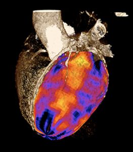
This image shows the areas of the heart receiving good blood flow (red) and other areas which are supplied by a blocked artery (darker colours).
This month two new papers were published in the Society of Cardiovascular Computed Tomography’s Journal which highlight the rapid advancement in medical technologies and the importance of staying at the forefront of imaging technology.
The papers describe a new way of imaging the heart called CT perfusion. Cardiac CT already allows the heart and the coronary arteries to be imaged in less than half a second with are resolution of half a millimetre or less. The technique has revolutionised the diagnosis of coronary disease as cholesterol build-up within the arteries (the cause of heart attacks) can be diagnosed without an invasive procedure and preventative treatment can be commenced.
For more complex situations or when a coronary stent or surgery is being considered information about blood flow to the heart muscle is also required. A CT perfusion study can assess both the extent of coronary blockages and the effect these have on flow to the heart muscle within a single visit.
Research led by Dr James Otton has shown that sophisticated four-dimensional processing algorithms result in significant improvements in CT perfusion image quality. In separate research, CT perfusion was compared with another emerging technique Cardiac MRI perfusion, also offered at St Vincent’s Hospital.
NSW Cardiology has a commitment to being at the forefront of heart care and treatment.
Links
A direct comparison of the sensitivity of CT and MR cardiac perfusion using a myocardial perfusion phantom.
Four-dimensional image processing of myocardial CT perfusion for improved image quality and noise reduction.

Recent Comments