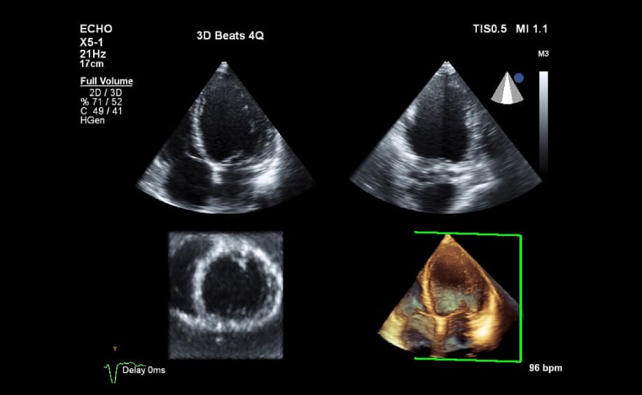
Echocardiography/ heart ultrasound is a non-invasive way of imaging the structure and function of the heart safely and painlessly. Using the latest ultrasound equipment we can build a detail picture of the heart chambers, major vessels, valves and blood flowing within the chambers in real time. These examinations may be at rest or after exercise on a treadmill.
New techniques such as three-dimensional echocardiography and strain analysis allow accurate measurement of heart volumes and contractility useful for the early detection of cardiomyopathy and coronary disease and is available in rooms.
You may be asked to have an ‘echo’/ultrasound to screen for signs and symptoms of heart problems, such as, heart failure, heart murmur or screening for congenital heart defects like ‘a hole in the heart’.
Your heart ultrasound can reveal;
How your heart muscle is pumping
The size of your heart
How your valves are working
If there is any fluid around your heart
If there are any blood clots inside your heart
If there are any problems with your heart’s major blood vessels such as your aorta
Cardio-Oncology
Chemotherapy and or radiotherapy can have adverse effects on the heart and circulation, during treatment and for years afterwards. Echocardiography plays an important role in closely monitoring the heart function whilst a patient is undergoing cancer treatment. We use the most up to date cardiac strain analysis tools to detect for early signs of heart failure.
Preparation
There is no preparation required for this test.
Procedure
An accredited Cardiac Sonographer will be performing the test. An instrument called a transducer will be placed in various positions on your chest. The transducer transmits and receives high frequency sound waves which transmits them as electrical impulses and converts them into moving pictures.
You will be required to be bare chested and will be given a gown to wear (front opening). Electrodes will be placed on your chest and you will be asked to roll on your left hand side to improve visualisation of your heart.
The transducer will be placed firmly on your chest to obtain images of your heart. You may hear noises that sound a bit like a washing machine, this is a magnified representation of blood flow through the heart valves and chambers.
You may be asked to take deep breaths in or hold a breath to get a better visualisation of your heart.
Scanning time will be approximately 40 minutes and may vary depending on the findings.
Results
A report will be generated by the Cardiologist, this will then go to your referring doctor usually the same day or overnight if done in the late afternoon. You can obtain your results from your referring Doctor or you can request from NSWC reception to send an official copy to you.
Have Questions?
Make an Appointment to get all your cardiology questions answered by our experienced team
Quick and Easy Consultation & Referral Process
